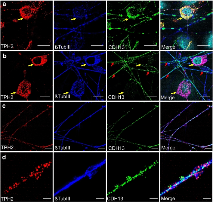
Fig. 4.
CDH13 expression in TPH2 + neurons using SIM. a IF staining of a TPH2 + neuron which is positive for CDH13 (yellow arrows). b IF staining of TPH2 + neurons which are negative for CDH13 (yellow arrows). Additionally, βTUBIII + fibers that are negative for TPH2 are immunoreactive for CDH13 and marked by a red arrow. c IF staining of TPH2 + fibers being positive for CDH13. d Close-up image of (c). Scale bar: a, b, c 10 μm; d 2 µm. Alexa Fluor 488 (βTUBIII), Alexa Fluor 555 (TPH2) and Alexa Fluor 647 (CDH13) were used to visualize target proteins

