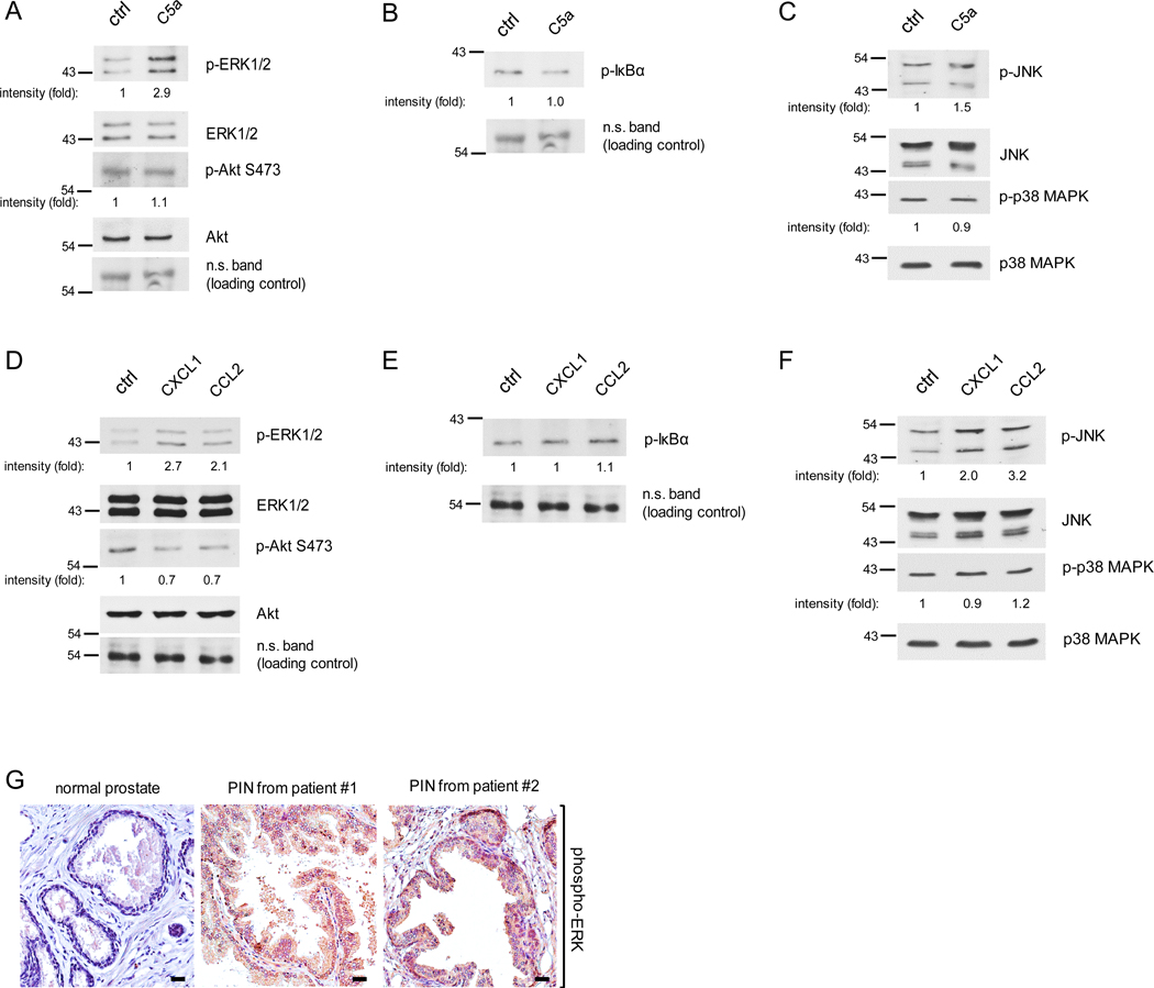
Figure 5. Activation of ERK and JNK in PIN cells stimulated by macrophage cytokines C5a, CCL2 or CXCL1.
(A-C) Pr111 cells grown on matrigel in 3D were treated with either control/ddH2O or recombinant C5a for 72h. Cell lysates were collected and subjected to immunoblotting for examining the protein of interests including p-ERK, ERK, p-AKt and Akt (A); p-IκBα (B); p-JNK, JNK, p-p38 MAPK and p-38 MAPK (C). (D-F) Similar to A-C, Pr111 cells cultured on matrigel in 3D were treated with control, recombinant CXCL1 or CCL2. Cell lysates were collected from 3D culture and subjected to immunoblotting for examining the levels of p-ERK, ERK, p-AKt and Akt (D); p-IκBα (E); p-JNK, JNK, p-p38 MAPK and p-38 MAPK (F). The relative intensity fold change was calculated by the ratio of phosphorylated protein levels over the corresponding total protein levels and set as 1 for control in each case. Each image shown in A-F was representative of 3–5 independent experiments. (G) Normal prostate and prostate cancer sample containing PIN of human tissues (n=3 per group) were immunostained for phosphorylated-ERK expression. Scale bar: 50 μm.

