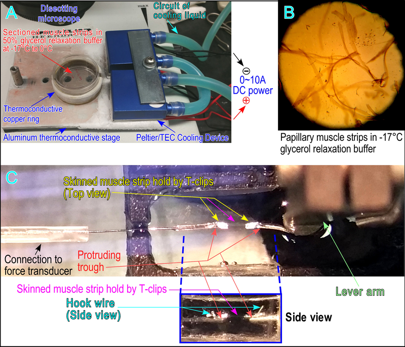
Figure 2. System set up for muscle strip selection, mounting and contractility measurements.
(A) A thermo-controlled aluminum stage was customer-made using two pairs of thermal electric coupler/Peltier with adjustable electric DC current to control the desired temperature on the surface of the aluminum plate. Heat produced by the Peltiers is removed by heat sinks connected to a water circulatory cooling system. The stage was placed under a dissection microscope with an underneath light source to luminate through the drilled holes. The muscle strips were placed in 50% glycerol relaxation buffer in a 35 mm cell culture dish with temperature controlled at 0°C. A thermo-conductive coper ring was attached to the culture dish to stabilize the temperature inside the dish. (B) Bright field view with underneath lightening visualizes the muscle strips under a dissecting microscope for selecting strip with properly organized myofibrils to mount to a pair of T-clips in 50% glycerol relaxation buffer at −17°C. (C) A skinned papillary muscle strip with T-clips was attached to metal wire hooks connected to force transducer and length-controlled lever arm. A pair of metal troughs (seen in the side view box) made from half-removed 30-gauge needles were glued underneath the hook as a support to prevent over-the-range movement of the T-clips when the muscle strip was lifted out of solution during the switch of buffer chambers.

