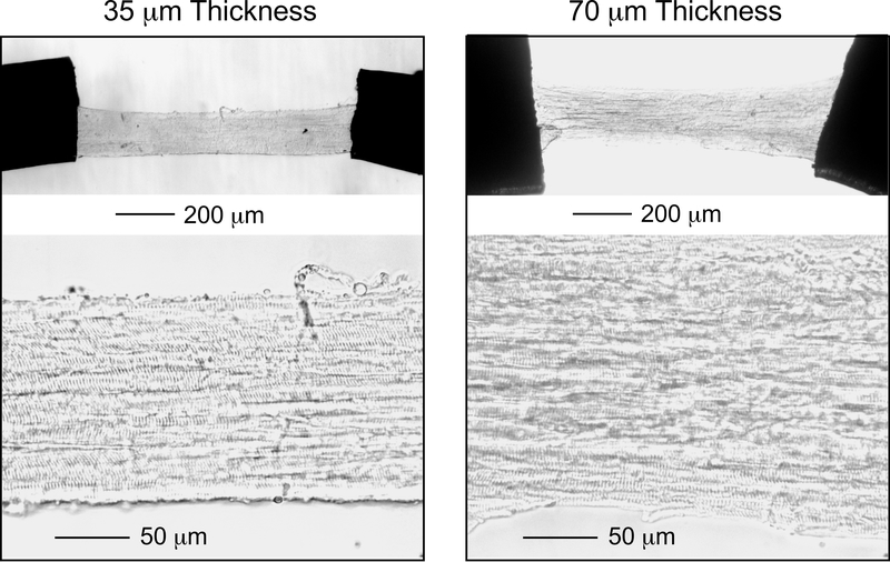
Figure. 3. Visual validation of the structure of 35 μm and 70 μm thick cryosection-generated skinned mouse left ventricular papillary muscle strips.
Papillary muscle strips with longitudinally and parallelly organized cardiomyocytes were selected and mounted via T-clips for contractility measurement (upper low magnification image). The lower left high magnification image of a 35 μm thick strip shows clear striations of skinned cardiomyocytes across the entire strip adjusted at a sarcomere length of 2.3 μm. The lower right image of a 70 μm thicker strip shows visible although less clear striations.

