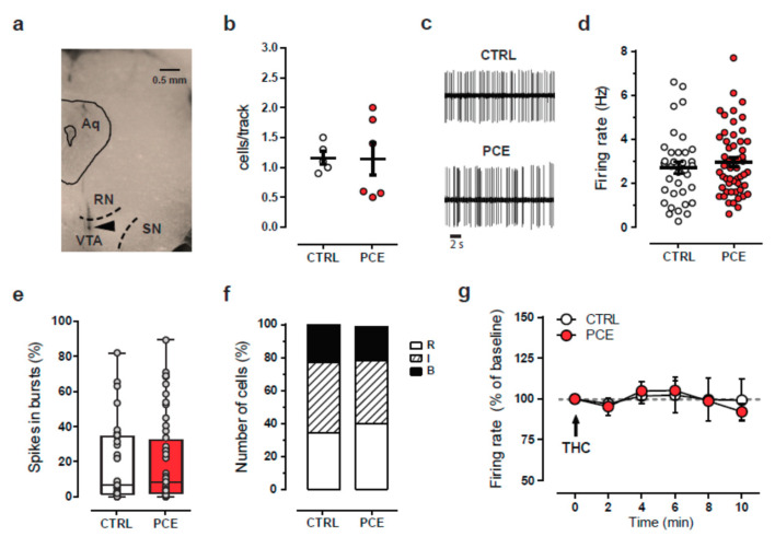
Figure 4.
PCE effect on electrophysiological properties of putative dopamine neurons recorded in vivo from female preadolescent rats. (a) Coronal midbrain section from a preadolescent female rat showing the recording site (black triangle) in the ventral tegmental area. Abbreviations: Aq, aqueduct; RN, red nucleus; SN, substantia nigra, VTA, ventral tegmental area. (b) Scatter plot showing the average number of spontaneously active VTA dopamine neurons encountered per track. PCE (nrats = 6) CTRL (nrats = 5). (c) Representative traces of spontaneous firing activity of dopamine neurons from female offspring. (d) Spontaneous firing frequency of VTA dopamine cells from CTRL and PCE female rats. Data are represented as means s.e.m. with single values. (e) Percentage of spikes in burst displayed by dopamine neurons in female offspring. Data are represented as a box-and-whisker plot with single values, min to max. (f) The stack bars represent the percentage of dopamine cells displaying different firing patterns: R = regular; I = irregular; B = bursty. (g) Time course of the effect of acute THC (0.5 mg/kg, i.v) on the firing frequency of putative VTA dopamine neurons from PCE (ncells = 5) and CTRL (ncells = 5). Data represented as average ± s.e.m.

