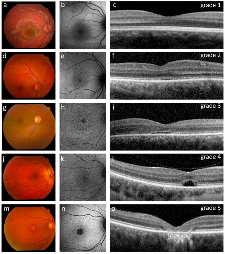
Figure 2.
Ophthalmological findings in five representative ACHM patients showing the OCT grading (c,f,i,l,o) with corresponding retinography (a,d,g,j,m) and fundus AF images (b,e,h,k,n). (a–c) Patient P15 (12 years old, homozygous for the c.619G>A variant in GNAT2) presented a normal fundus appearance, normal fundus autofluorescence and a grade 1 OCT with a continuous ellipsoid zone (EZ band). (d–f) Patient P21 (16 years old) had a normal appearing fundus, reduced autofluorescence with subtle hyper-autofluorescence around the nasal fovea, and a grade 2 OCT with a disrupted EZ band. (g–i) Patient P19 (19 years old) presented a normal fundus, foveal and parafoveal hyper-autofluorescence, and a grade 3 OCT characterized by an absent EZ band. (j–l) Patient P6 (32 years old, homozygous for the c.847C>T variant in CNGA3) had a normal fundus appearance, foveal and parafoveal hyper-autofluorescence, and a grade 4 OCT characterized by the presence of a hypo-reflective zone. (m–o) Patient P10 (46 years old, homozygous for the c.1148delC variant in CNGB3) presented retinal pigment epithelium (RPE) dystrophy, a central region devoid of autofluorescence surrounded by a hyper-autofluorescent ring, and a grade 5 OCT characterized by outer retinal atrophy including RPE loss.

