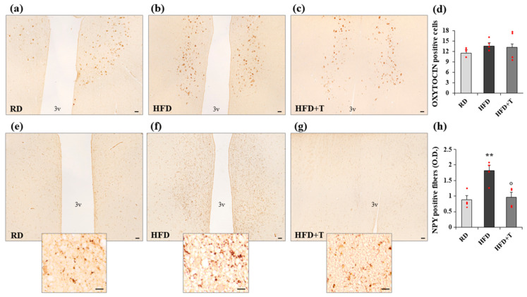
Figure 5.
Immunostaining of oxytocin and neuropeptide Y (NPY) positivity in the hypothalamic paraventricular nucleus (PVN) from RD, HFD, and HFD + T rabbits. Representative images of coronal sections showing the presence of both oxytocin-positive neurons (a–c) and NPY-positive fibers (e–g, higher magnification in the inset) localized in the PVN, adjacent to the third ventricle (3v). (d) Quantification of oxytocin-positive neurons obtained by counting 10 fields in four different samples from each group in the PVN area (means ± SEM, n = 4 for each group); (h) Computer-assisted analysis of NPY-positive fibers, calculated in 10 fields of four different samples from each group (mean ± SEM, n = 4 for each group; ** p < 0.01 vs. RD; ° p < 0.05 vs. HFD). Scale bar = 50 μm (lower magnification) and 10 μm (higher magnification).

