Figure 2.
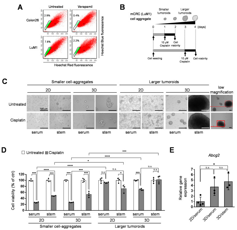
Tumoroids acquired platinum-resistance in metastatic colorectal cancer (mCRC) cells: (A) analysis of side population (SP) cells. Colon26 or LuM1 cells were treated with Hoechst 33342 and/or verapamil for flow cytometry. Green dots in the enclosed area indicate SP cells, whereas red dots are the main population cells; (B) schemes of the experimental protocol. Smaller cell aggregates were formed by pre-culturing LuM1 cells for 1 day. Larger tumoroids were formed by pre-culturing the cells for 3 days. Cisplatin at 10 μM was added to the tumoroids, and cell viability was measured; (C) representative images of cells and tumoroids cultured in 2D vs. 3D culture plates. Cells were cultured in a serum-containing medium or mTeSR1 stem cell medium. Scale bars, 100 μm; (D) cell viabilities of LuM1 cells and tumoroids treated with Cisplatin or untreated. n = 3 or 4 independent culture wells; (E) gene expression of Abcg2 in 2D vs. 3D culture conditions. n = 3 or 4 independent culture wells. * p < 0.05, *** p < 0.001, **** p < 0.0001, n.s., not significant.

