Figure 3.
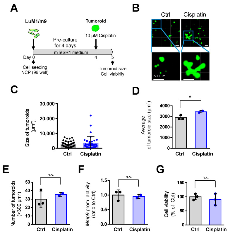
Cisplatin (at 10 μM) promoted the tumoroid growth of mCRC cells: (A) a scheme of the experimental protocol used for panel B–G. LuM1/m9 cells were pre-cultured for 4 days in mTeSR1 on NanoCulture Plate® (NCP), and then Cisplatin at 10 μM was added; (B) representative images of tumoroids. Scale bars, 500 μm; (C) column scatters the plotting of tumoroid size. Representative data from a single well were shown; (D) average tumoroid size. n = 2 or 3 independent culture wells; (E) the number of tumoroids larger than 300 μm2. n = 2 or 3 independent culture wells; (F) Mmp9 promoter activity. n = 3 independent culture wells; and (G) cell viability. n = 3 independent culture wells. * p < 0.05, n.s., not significant.

