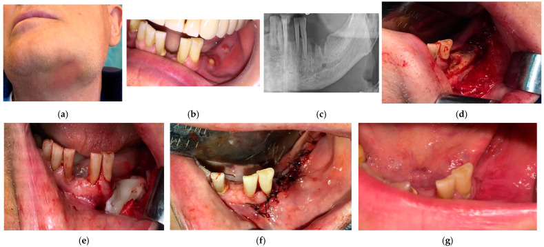
Figure 6.
(a) Patient in stage 3 of MRONJ with infection extended to the inferior border of mandible-extraoral view; (b) intraoral view at the initial presentation of the patient; (c) mandible bone showing a sclerotic structure; (d) performed surgery, with necrotic block resection; (e) PRF membranes covering completely the bone in 2 layers; (f) tension-free closure of the surgical wound; (g) complete healing achieved 6 months after the end of the treatment.

