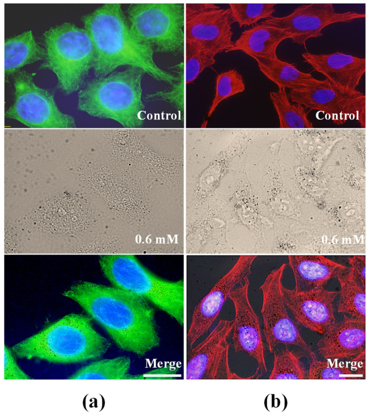
Figure 6.
Analysis of cytoskeleton in HeLa cells incubated 24 h with 0.6 mM metal concentration and observed under fluorescence and bright-field microscopy immediately after incubations. (a) Immunofluorescence staining of α-tubulin observed under blue light excitation. (b) TRITC-phalloidin visualization of actin microfilaments observed under green light excitation.

