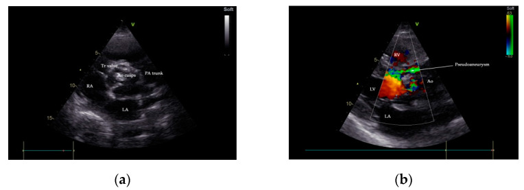
Figure 2.
Transthoracic echocardiography (TTE), parasternal short-axis-base view, showing a heavily calcified aortic valve with possible vegetation (a); communication between LVOT and P-MAIVF is shown by color flow Doppler, parasternal-long axis view (b). Ao—aorta, LA—left atrium, LVOT—left ventricular outflow tract, PA—pulmonary artery, P-MAIVF—pseudoaneurysm of the mitral-aortic intervalvular fibrosa, RA—right atrium, RV—right ventricle, Tr—tricuspid valve.

