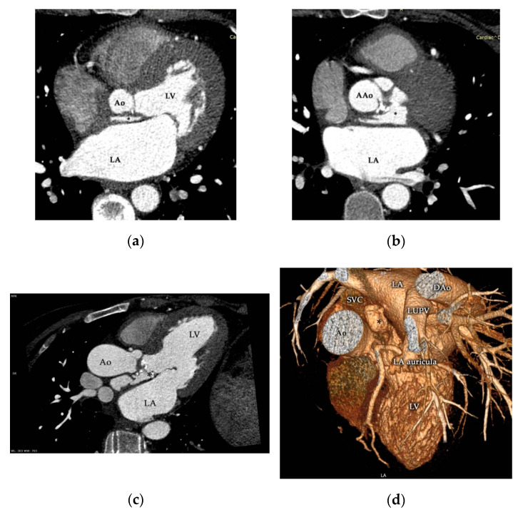
Figure 5.
Contrast-enhanced ECG-gated multidetector-row cardiac computed tomography (MDCT) study of the aortic valve and ascending aorta during the cardiac cycle corroborated the presence of P-MAIVF (*) with the appearance of contrast media (a–c). Three-dimensional reconstruction (d) supported the aforementioned findings, and precise localization is clearly demonstrated. Ao—aorta, AAo—ascending aorta, DAo—descending aorta, LA—left atrium, LUPV—left upper pulmonary vein, LV—left ventricle, SVC—superior vena cava.

