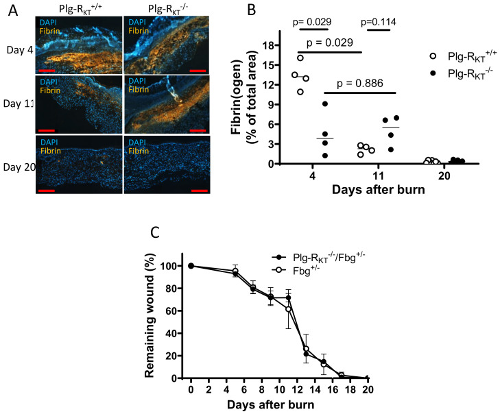
Figure 3.
Enhanced fibrin(ogen) content in Plg-RKT−/− wound tissue and effect of fibrinogen heterozygosity on burn wound healing. (A) Immunohistochemical staining for fibrin in the tissue of Plg-RKT−/− and Plg-RKT+/+ mice collected at different time points after induction of burn wounds (n = 4). Scale bar = 200 µm. (B) Quantification of fibrinogen area in Panel A (%) based on immunostaining using Image J software https://imagej.nih.gov/ij/download.html. (C) Quantification of the remaining wound area at different time points after burn wounding of male Plg-RKT−/−/Fgn+/− (n = 4) mice and Plg-RKT+/+/Fgn+/− mice (n = 5). Post hoc testing was done by Mann–Whitney test. This figure is modified from a figure originally published in [80]. Creative common license available at: http://creativecommons.org/licenses/by/4.0/.

