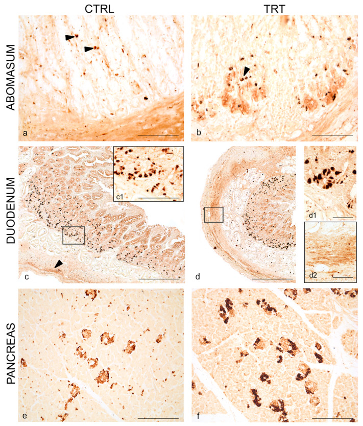
Figure 1.
Neuropeptide Y (NPY) immunoreactivity in the abomasum, duodenum and pancreas of control and treated goat kids. Scattered positive cells (arrowheads) in the epithelium of gastric mucosa of control (a) and anthocyanins (ANTs) fed animals (b). Overview of NPY distribution in neuroendocrine cells (c) in the crypt of Lieberkühn and varicose positive fibers (arrowhead) in the muscular layer of duodenum of control (a) and ANTs fed animals (d). Higher magnification of neuroendocrine cells in the crypt of Lieberkühn (c1,d1) and fibers in the muscular layer (d2). Overview of NPY distribution in the pancreatic islets of control (e) and ANTs fed animals (f). Scale bar: (a,b,e,f) = 50 µm; (c,d) = 100 µm; (c1,d1,d2) = 25 µm.

