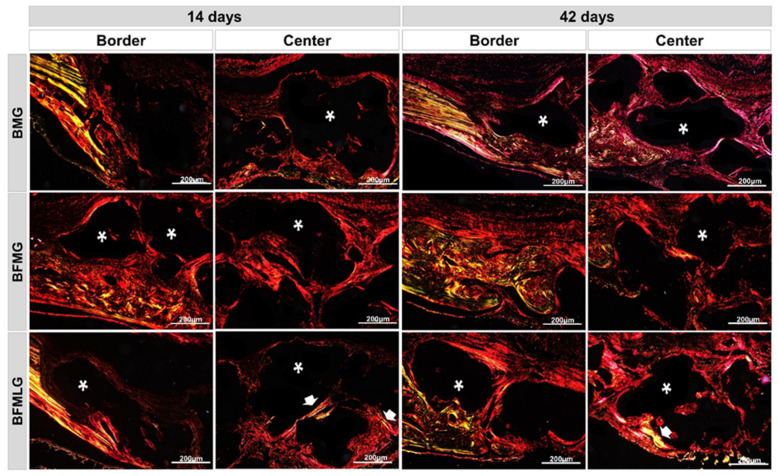
Figure 6.
Histological sections of the border and center of the bone defect of rat calvaria stained by Picrosirius-red under polarized light at 14 and 42 days. BMG—defects filled with HA/β-TCP covered by a bovine biological membrane; BFMG defects filled with HA/β-TCP and heterologous fibrin biopolymer covered with bovine biological membrane; BFMLG defects filled with HA/β-TCP and heterologous fibrin biopolymer covered with bovine biological membrane and PBMT. RGB green-yellow-red colors. Mature bone—yellowish-green fibers (white arrow); immature bone—orange-red; particles of the biomaterial (dark background—asterisk). Original 10× magnification, 200 µm scale bar.

