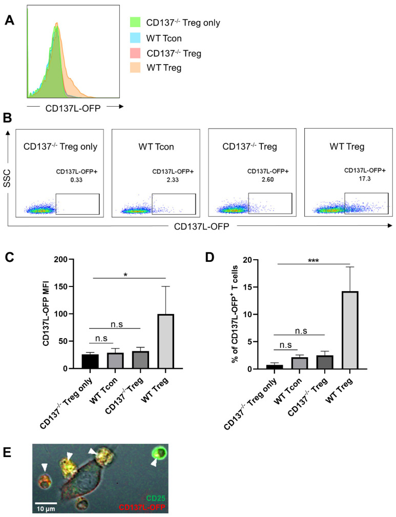
Figure 2.
WT Treg are most effective in depleting CD137L from APC. Sorted WT Treg or WT Tcon or CD137−/− Treg were cocultured with RAW-CD137L-orange fluorescent protein (OFP) cells for 1 h at a ratio of 1:1. (A) Cells were gated for live and single CD4+ cells, and CD137L-OFP levels on T cells were determined by flow cytometry. (B) Representative dot plots of the CD137L-OFP signal in CD137−/− Treg before the coculture and in WT Treg, WT Tcon, and CD137−/− Treg after the coculture. The number above the gate represents the percentage of the CD137L-OFP+ population. (C) MFI of CD137L-OFP of CD137−/− Treg before the coculture and of WT Treg, WT Tcon, and CD137−/− Treg after the coculture. (D) Percentage of CD137−/− Treg before the coculture and of CD137L-OFP+ WT Treg, WT Tcon, and CD137−/− Treg after the coculture. Data are representative of three independent experiments. Graphs show means ± SD, * p < 0.05, *** p < 0.001, n.s. not significant, one-way ANOVA with Dunnett multiple comparison test. (E) Confocal image of a 24 h coculture of CD25+ WT Treg and RAW-CD137L-OFP cells. Yellow spots indicate the internalization of CD137L-OFP (red) into CD25+ Tregs (green). White arrows indicate the location of the transferred CD137L-OFP in Treg.

