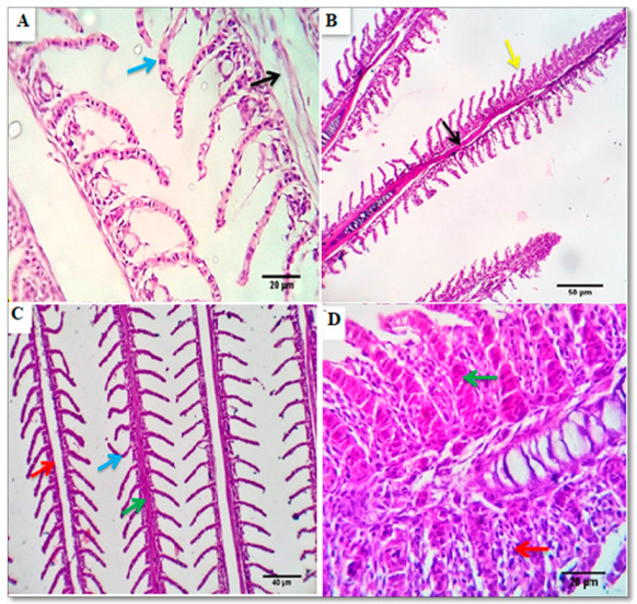
Figure 3.

Photomicrograph from gills the control group (A) showing normal primary (black arrow) and secondary gill filaments with normal pavement cells, (blue arrow) Scale bars, 20 μm. (B) photomicrograph from the gills of the BSRE5 group, showing tips of a few filaments with epithelial lifting (yellow arrow) and stromal lymphocytic infiltration, Scale bars 50 μm. (C) photomicrographs from the gills of the BSRE10 group showing congested, Telangiectatic capillaries (green arrow), focal denudation of the secondary filament structures (blue arrow), lymphocytic infiltration (red arrow). Scale bars, 40 μm. (D) photomicrographs from the gills of the BSRE15 group showing that the tips of some filaments appear focally denuded, fused, thick, and enlarged by severely congested capillaries (green arrow), epithelial proliferation, goblet cell proliferation, and lymphocytic infiltration (red arrow).
