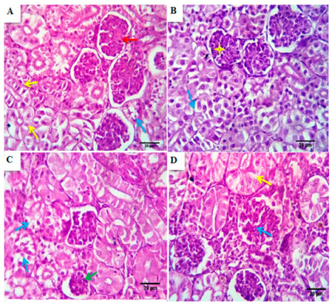
Figure 5.

Photomicrographs from the kidney of the control group (A) showed normal renal glomerular, tubular, and interstitial structures with preserved Bowman’s capsular histomorphology, glomerular capillary morphology (red arrows), and tubular epithelial length and widths, minimal degenerative changes in a few numbers of renal tubules were seen (yellow arrows), Scale bar, 20 μm. (B–D) Photomicrographs from the kidney of BSRE5, BSRE10, and BSRE15 groups, respectively, showed comparatively healthy, normal counterparts of the nephron units with a slandered morphological appearance, which is homologous to the control one in BSRE5 group (blue arrows and yellow stars) and less standardized in BSRE10 and BSRE15 groups, where some of the renal tubules suffered degenerative changes, mostly of hydropic type ((C), blue arrows), ((D), yellow arrows), Scale bar, 20 μm.
