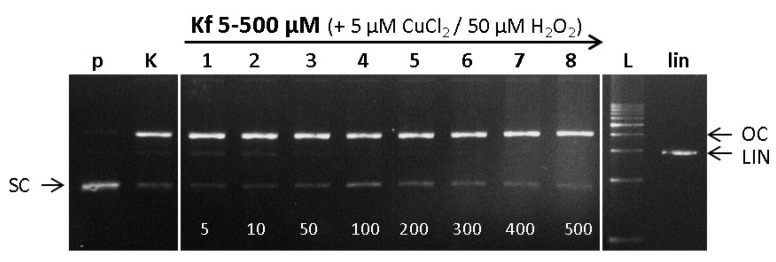
Figure 7.
Electrophoretic profile of agarose gel (0.8%), 15 µM plasmid PBSK + DNA with Cu(II) ions, hydrogen peroxide and kaempferol incubated for 30 min. Samples were loaded in the gel lanes in the following order: (p) pDNA–control; (K) pDNA + 5 µM CuCl2 + 50 µM H2O2-control Cu-Fenton reaction; (1–8) pDNA + 5 µM CuCl2 + 50 µM H2O2 + (1) 5 µM kaempferol, (2) 10 µM kaempferol, (3) 50 µM kaempferol, (4) 100 µM kaempferol, (5) 200 µM kaempferol, (6) 300 µM kaempferol, (7) 400 µM kaempferol, (8) 500 µM kaempferol; (L) 1 kb DNA standard, (lin) pDNA linearized with EcoR1 endonuclease.

