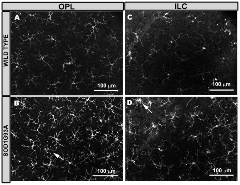
Figure 1.
Microglial cells in the outer plexiform layer (OPL) and in the inner layer complex (ILC) constituted by the inner plexiform layer and nerve fiber layer–ganglion cell layer. Retinal whole-mount was labeled with anti-Iba-1. In aged-matched wild type mice, the microglial cells of the OPL (A) and the ILC (C) showed a ramified morphology, featuring primary processes from which they derive secondary processes, and constituted a regular plexus of tiled cells along the retina. ILC microglia processes were thinner than those of OPL. In SOD1G93A mice, the microglia of the OPL (B) and the ILC (D) showed thickening of the cell body and processes, giving the cell a more robust and larger appearance. Some microglial cells showed a retraction of these processes (arrow). Number of retinas used in the experiment, WT: n = 6; and SOD1G93A: n = 6.

