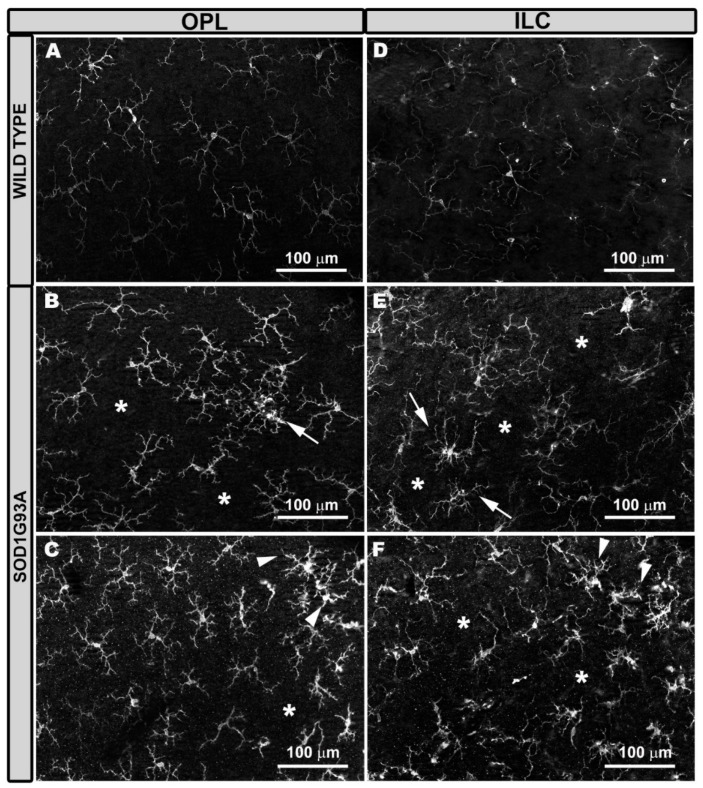
Figure 2.
Microglial cells in the outer plexiform layer (OPL) and inner layer complex (ILC) constituted by the inner plexiform layer and nerve fiber layer–ganglion cell layer. Retinal whole-mount labeled with anti-Iba-1. Compared to wild type mice (A,D), in SOD1G93A mice the microglial plexus was not as regular in OPL (B,C) and in ILC (E,F). There were areas where the microglia grouped together and featured retracted processes, leaving areas without cells (*). The groups of cells were formed either in circles (B,E) (arrows) or in rows (C,F) (arrowheads). Number of retinas used in the experiment, WT: n = 6; and SOD1G93A: n = 6.

