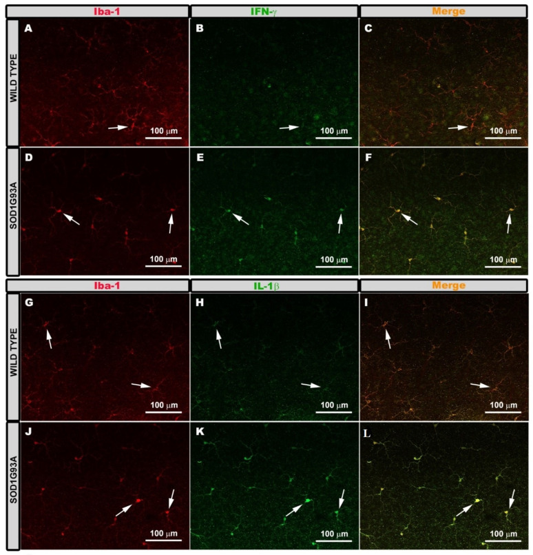
Figure 3.
Pro-inflammatory M1 phenotype. Retinal whole-mounts are labeled with anti Iba-1 and anti IFN-γ (A–F) and with anti-iba-1 and anti-IL-1β (G–L) showing the microglial plexus in the outer plexiform layer. Immunoreactivity for IFN-γ (A–C) and IL-1β (G–I) in the Iba-1+ cells was very low in the wild type group (arrow). Iba-1+ cells of the SOD1G93A group showed very intense immunoreactivity for IFN-γ (D–F) and IL-1β (J–L) (arrows). Number of retinas used in the experiment, WT: n = 3; and SOD1G93A: n = 3.

