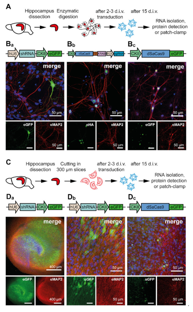
Figure 3.

AAV-mediated expression of different constructs in primary hippocampal neurons (PHNs) and organotypic hippocampal slice cultures (OHSCs). (A) Schematic representation of the preparation and transduction procedure of primary hippocampal neurons (PHNs). (Ba–Bc) Representative immunofluorescence images of rAAV9-transduced PHNs expressing the (Ba) eGFP reporter, (Bb) HA-tagged dSaCas9, and (Bc) eGFP-tagged dSaCas9. (C) Schematic representation of the preparation and transduction procedure for OHSCs. For details see Material and Methods. (Da–Dc) Representative immunofluorescence images showing rAAV9-transduced OHSCs expressing the eGFP reporter (Da,Db), or eGFP-tagged dSaCas9 (Dc). The eGFP, HA-tag, and the neuron-specific microtubule-associated protein 2 (MAP2) protein were immunostained with specific anti-GFP, anti-HA, and anti-MAP2 antibodies combined with fluorescently labeled secondary antibodies (eGFP and HA-tag, green; MAP2, red). Nuclei were labeled with TOPRO (blue). Schematic of AAV-delivered constructs are displayed above the merged immunofluorescence images.
