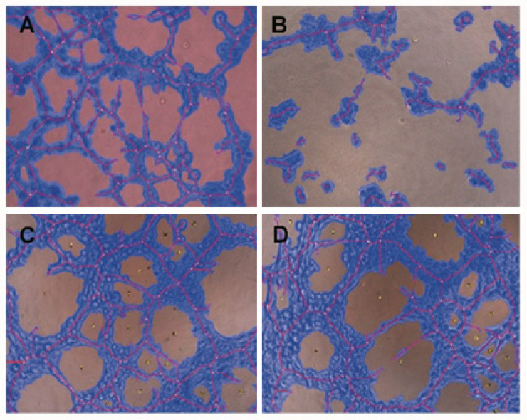
Figure 4.
Capillary- like structures formed by HUVEC cells after incubation with conditioned medium from control cells (A); SDC-1 over-expressing cell (B), control medium from scrambled control cells (C), and MMP7 silencing cell medium (D). Images were taken with 10X magnification. Red line defines the tube’s length and blue color shows capillary structures. Small yellow dots demonstrate the branching points.

