Figure 6.
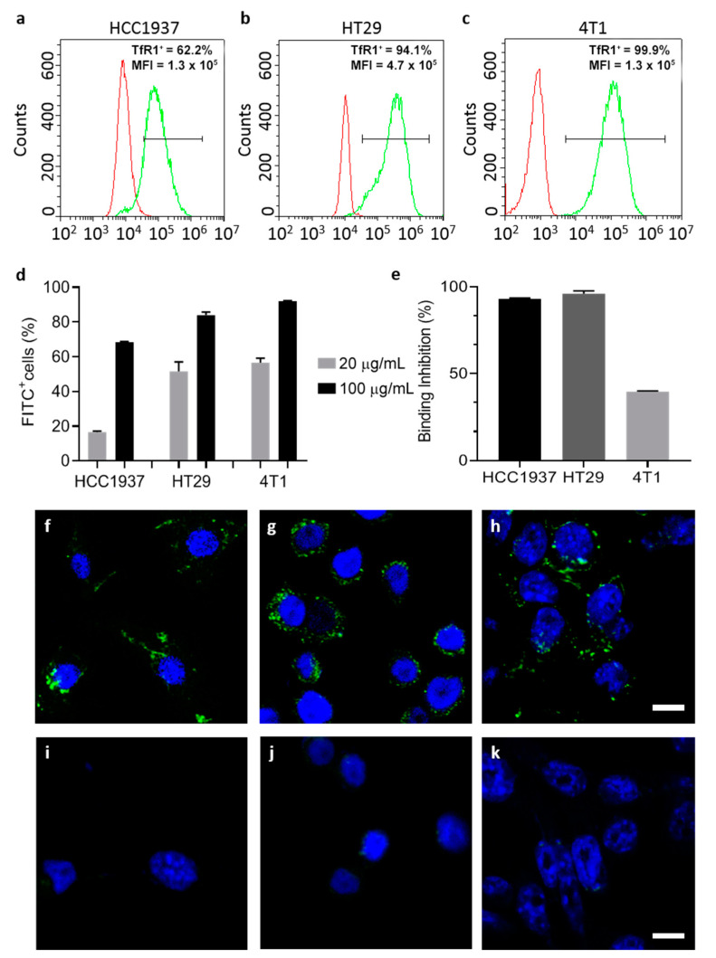
Cell interactions and uptake of ETX-free HFn. Representative histograms of TfR1 expression of selected cells (HCC1937, HT29 and 4T1) studied by flow cytometry (a–c respectively) (TfR1+: percentage of TfR1 positive cells, after setting proper fluorescence fates on non-stained cells; MFI: mean fluorescence intensity). Cells have been incubated with F-HFn at different concentrations at 4 °C for 2 h and binding has been evaluated by flow cytometry showing that binding is correlated with TfR1 expression (d); competition assay showed reduced binding of HFn FITC when cells were pre-incubated with non-labelled HFn, confirming the TfR1 binding specificity (e); results in panels d and e are reported as average ± S.D. of 3 independent experiments. Confocal microscopy micrographs showing uptake of ETX-free F-HFn in HCC1937, HT29 and 4T1 after incubation with 100 µg/mL of particles for 6 h (f–h). Cells were incubated with the same concentration of free FITC for 6 h and the signal was almost undetectable (i–k) F-HFn and FITC are represented in green, while nuclei labelled by DAPI are colored in blue. Scale bar 10 µm.

