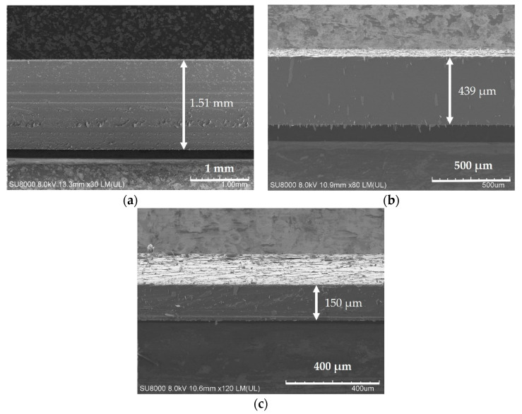
Figure 2.
Scanning electron microscope (SEM) micrographs of the cross-sectional morphology of the PDMS substrates prepared with the three different spinning speeds. (a): The cross-sectional of PDMS thickness with 1.51 mm at the rotation speed of 0 r/min; (b): The cross-sectional of PDMS thickness with 439 μm at the rotation speed of 500 r/min; (c): The cross-sectional of PDMS thickness with 150 μm at the rotation speed of 1000 r/min.

