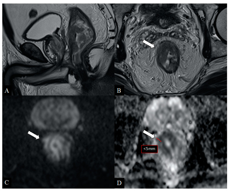
Figure 3.
48-year-old patient with rectal cancer. Sagittal (A) and axial (B) T2-weighted images show heterogeneous tissue involving the right hemicircumference of the lower rectum with a focal breakpoint at 10 o‘clock (arrow) doubtful due to wall infiltration (T2/T3 stage). The diffusion-weighted axial images (C) with relative ADC map (D) show a clear extraparietal component < 5 mm (arrows) with diffusion restriction and very low ADC map values (pathological hypercellular tissue). Histological examination confirmed the mesorectal extension of the disease (stage T3b).

