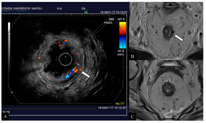
Figure 4.
Early-stage tumor affecting the middle rectum in a 64-year-old woman. Endorectal ultrasound (ERUS) scan (A) demonstrates the presence of pathological rectal tissue confined to the left submucosal wall with preserved definition of the hypoechoic layer of muscolaris propria (arrow) referable to an initial clinical stage (T2) of the disease. The axial T2-weighted images (B,C) obtained with magnetic resonance imaging (MRI) raised the suspicion of extra-parietal infiltration due to evidence of spiculation in the perivisceral mesorectal tissue (arrow). The histological examination confirmed the desmoplastic nature of the tissue with the presence of granulocytes and eosinophils, suggestive for a reactive/inflammatory tissue.

