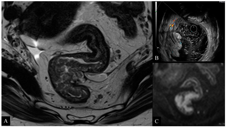
Figure 5.
67-year-old woman with high rectal cancer. Axial T2-weighted image (A) obtained with MRI shows a large circumferential tumor infiltrating the muscolaris propria layer and close to the anterior peritoneal reflection. ERUS scan (B) and axial diffusion weighted imaging (DWI) (C) images confirm the presence of a tumor infiltrating the rectal wall with focal invasion to the perirectal fat. Pathology examination confirmed the presence of a T3b tumor.

