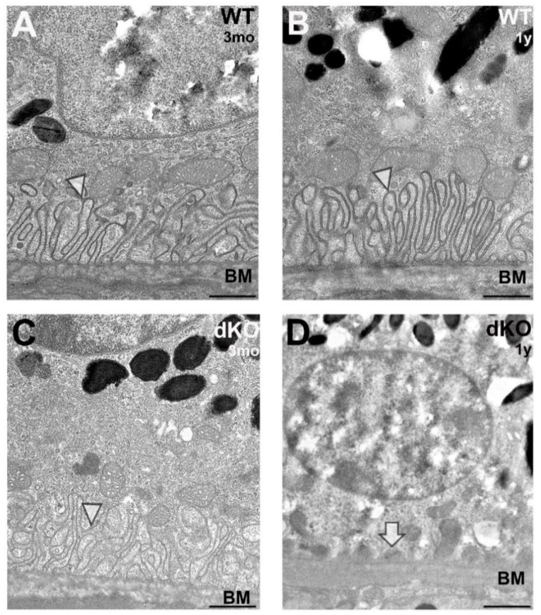
Figure 3.
Structural changes of the basal compartment of the retinal pigment epithelium (RPE) in NFE2L2/PGC-1α double knockout (dKO) mice as compared to their age-matched wild-type (WT) counterparts. Representative high power transmission electron microscopy (TEM) images of the samples of three-months- (A,C) and one-year-old (B,D) WT and dKO animals are shown. RPE of WT mice at both time points (A,B) and the three-months-old dKO samples (C) show normal basal arrangement: well-organized infoldings above Bruch’s membrane, supporting intracellular organelles (infoldings indicated by arrowheads) and typical thickness of Bruch’s membrane (BM). In contrast, many of the one-year-old NFE2L2/PGC-1α dKO mice RPEs show ultrastructural signs of loss of the basal infoldings with basal linear deposits (D; arrow), resulting in unsupported intracellular organelles above Bruch’s membrane. Scale bars: 1 µm.

