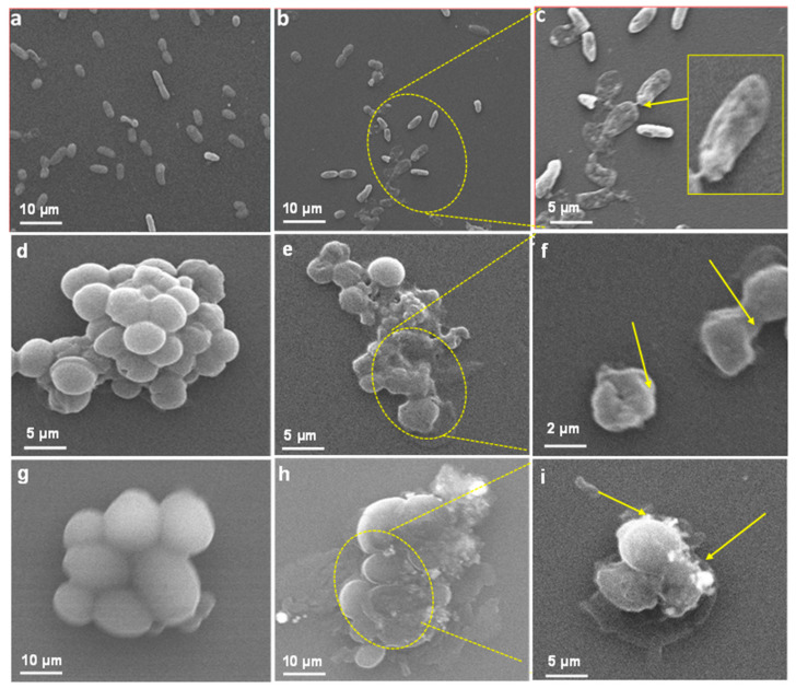
Figure 8.
Interaction between LIV-AgNPs and microbial cells. Panels (a,d,g) represent SEM images of MDR-PA, MRSA and C. albicans cells in the absence of PLE-AgNPs. Whereas, panels (b,c,e–i), demonstrate cellular damage in MDR-PA, MRSA and C. albicans cells in presence of 100 µg/mL of LIV-AgNPs at low (10–5 µm) and high (5–2 µm) scales, respectively.

