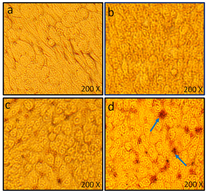
Figure 12.
Microscopic analysis of HCT-116 cells exposed to varying concentrations of LIV-AgNPs for 24 h. Cytotoxic effects were manifested as shrunken morphology, gaps between the neighboring cells and cellular detachments, which appeared as round bodies in the culture medium. Images were captured at the magnification of 200X using a bright field inverted microscope (Olympus, CKX41, Tokyo, Japan). (a) HCT-116 untreated cells, (b–d) HCT-116 cells treated with—10, 50 and 100 µg/mL of LIV-AgNPs, respectively.

