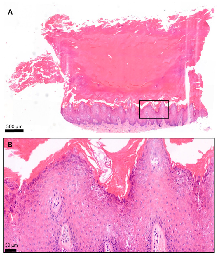
Figure 8.
Histology of the plantar keratoderma of the patient, hematoxylin and eosin (H&E) staining. The skin biopsy specimen had a structure typical of acral localization. (A) The surface was covered by a very thick hyperkeratotic and orthokeratotic horny layer. (B) The epidermis was significantly acanthotic and slightly papillomatous. The stratum granulosum was widened. The keratohyaline granules were differently shaped and sized and showed perinuclear vacuolization of the keratinocytes. The basal membrane was intact. (A) 2.8× magnification; scale bar: 500 µm; pixel size: 0.184 pixels/µm. (B) 19.9× magnification; scale bar: 50 µm; pixel size: 1.12 pixels/µm.

