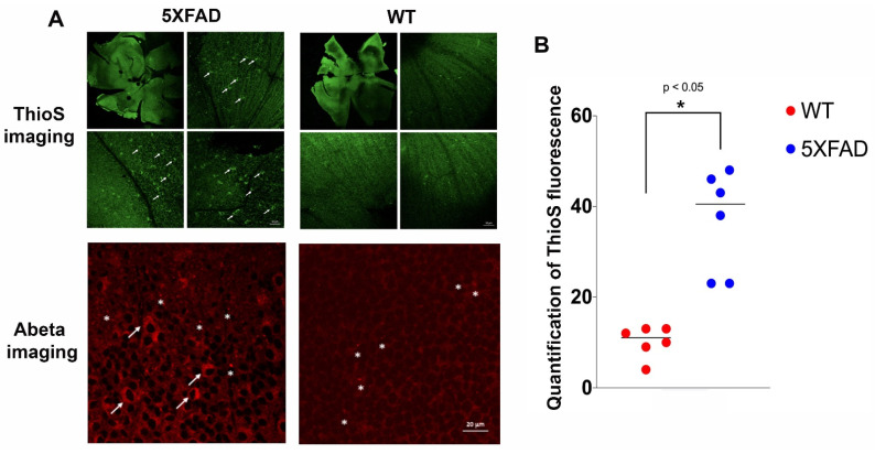
Figure 2.
Representative images of confocal fluorescence imaging of retinal whole-mount of treated wild type (WT) versus 5XFAD mice. (A) ThioS fluorescence (green, 488 nm channel) are prominent in different areas of the 5XFAD retinae compared to those from WT counterparts. There are some signals in the WT retinae, although much less, but detectable. Selected areas are magnified to show more detailed information. The fluorescence signal appears in the intracellular compartment of the retinal ganglion cells (RGC, arrows). These results corroborate with the retinal amyloid beta (Abeta) immunohistochemistry (red, 546 nm channel) shown in the bottom panels in which RGCs are labeled in 5XFAD (arrows) and extracellular Abeta deposits are also present (asterisks) in both 5XFAD and WT mice; (B) quantification of the ThioS signal was performed on retinal sections from WT and 5XFAD mice, and the signal distribution was scored on an ordinal scale after thresholding using Otsu method, and presented in the vertical scatterplot. An asterisk indicates significant differences between WT vs. 5XFAD (p < 0.05).

