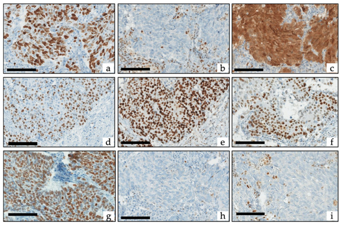
Figure 1.
Example of Ki-67, retinoblastoma protein (Rb), p16 and p53 immunohistochemistry: (a–c) SCNEC with (a) Ki-67 at 90%, (b) loss of Rb (with positive internal controls) corresponding to the absence of Rb staining (Rbinap) pattern, and (c) high expression of p16 corresponding to the intense p16 staining (p16high) pattern. (d–f) LCNEC with (d) Ki-67 at 60%, (e) diffuse overexpression of p53 corresponding to the inappropriate p53 staining (p53inap) pattern, and (f) conserved expression of Rb corresponding to the appropriate Rb staining (Rbapp) pattern. (g–i) SCNEC with (a) Ki-67 at 83%, (h) complete loss of p53 (with positive internal controls) corresponding to the p53inap pattern, and (i) loss of Rb (with positive internal controls) corresponding to the Rbinap pattern. Scale bars correspond to 50 µm.

