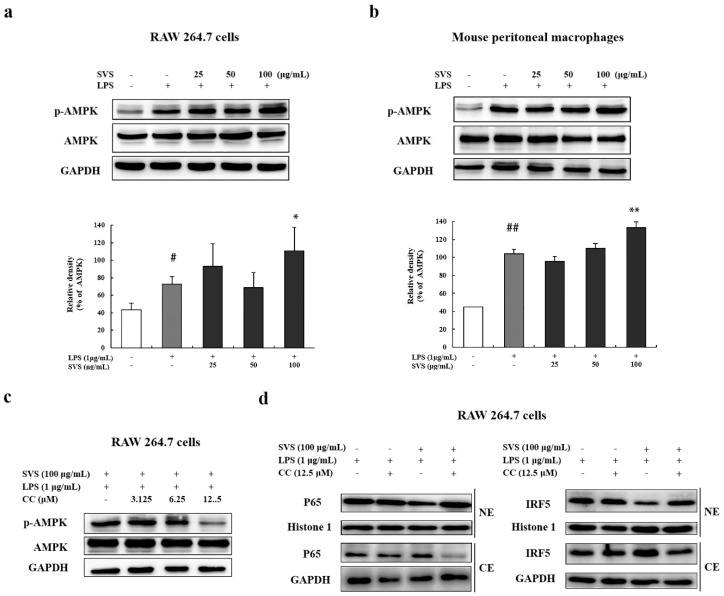
Figure 8.
Stevioside inhibited the IRF5/NF-κB pathway through AMPK activation. (a,b) Protein levels of AMPK and phosphor-AMPK (p-AMPK) were detected by Western blot in RAW 264.7 cells and mouse peritoneal macrophages, which were pretreated with or without stevioside (25, 50, or 100 μg/mL) for 1 h and then stimulated with LPS (1 μg/mL) for 1 h. The lower panels are quantifications of the blots, as measured by Image J; (c) Protein levels of AMPK and phosphor-AMPK (p-AMPK) were examined by Western blot in RAW 264.7 cells treated with indicated concentrations of compound C (CC) for 1 h, followed by 100 μg/mL stevioside treatment for 1 h and 1 μg/mL LPS stimulation for another 1 h; (d) Protein levels of P65 (left panel) and IRF5 (right panel) were detected by Western blot in nuclear (NE) and cytosolic extracts (CE) of RAW 264.7 cells, which were treated as in (c) using 12.5 μM CC. Histone 1 and GAPDH were used as internal controls. # p < 0.05, ## p < 0.01 versus control group; * p < 0.05, ** p < 0.01 versus LPS-treated group. SVS, stevioside.

