Figure 7.
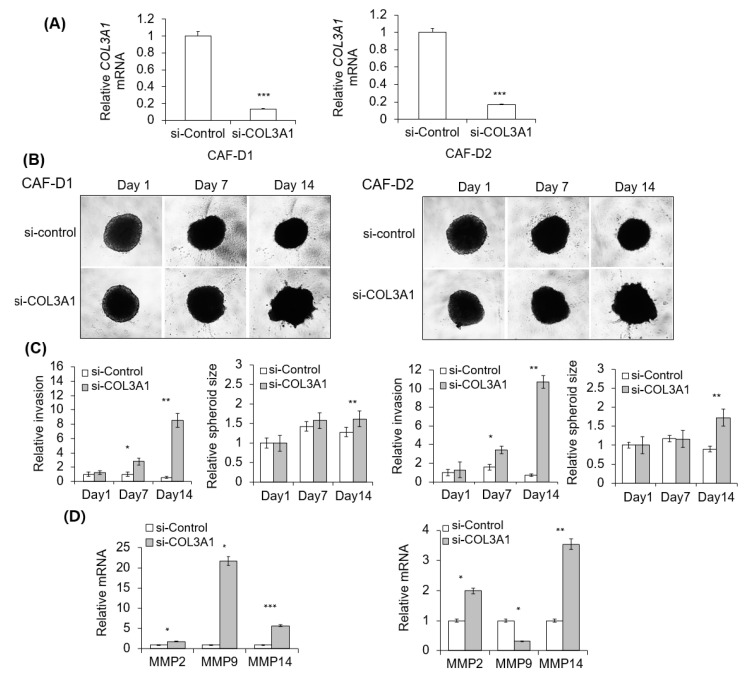
Effect of collagen knockdown-CAF-D on FaDu spheroid invasion. (A) CAF-D cells were transfected with siCOL3A1 for two days, and CM was collected. siRNA efficiency was evaluated using qPCR analysis. (B) FaDu spheroid (400 μm in diameter) was formed by culturing a 96-well U-bottom Ultra-Low Attachment plate for two days. CM and Matrigel were added to each well, and the spheroid invasion was monitored for 14 days using phase-contrast microscopy (5× magnification). (C) Cell invasion was quantified by measuring the mean number of tube-like structures extending from the surface of each spheroid. The spheroid size was quantified using Cell3iMager. (D) MMPs mRNA expression in FaDu spheroids were analyzed using qPCR on day 14. Results represent the mean ± standard deviation of three experiments (* p < 0.05, ** p < 0.01, *** p < 0.005).

