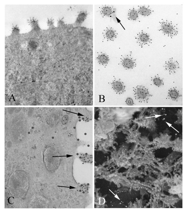
Figure 1.
Co-localization of respiratory syncytial virus (RSV) and nucleolin in infected MDCK cells: (A) TEM of an RSV infected MDCK cell. Note the virus particles associated with the apical surface. Magnification 60,000×. (B) Cross sections of virus particles near the apical surface which have been double immunogold labeled for RSV (small particles) and nucleolin (large particles). A small portion of the apical membrane is seen with the virus, which is labeled with large particles (arrow). Magnification 60,000×. (C) TEM of a RSV infected MDCK cell, immunogold labeled for nucleolin and RSV. Nucleolin is seen at the periphery of the viral particles associated with the apical membrane (arrows) (large particles), while RSV antibody label (small particles) was confined to the interior of the virus. (D) SEM of the surface of an infected cell was imaged using a Wein filter. Numerous co-localizations are seen (arrows). Magnification 70,000×.

