Figure 3.
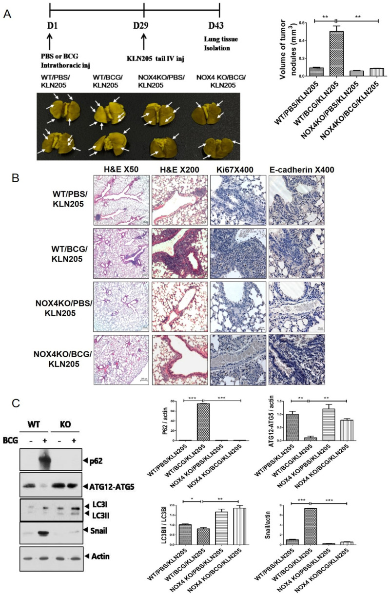
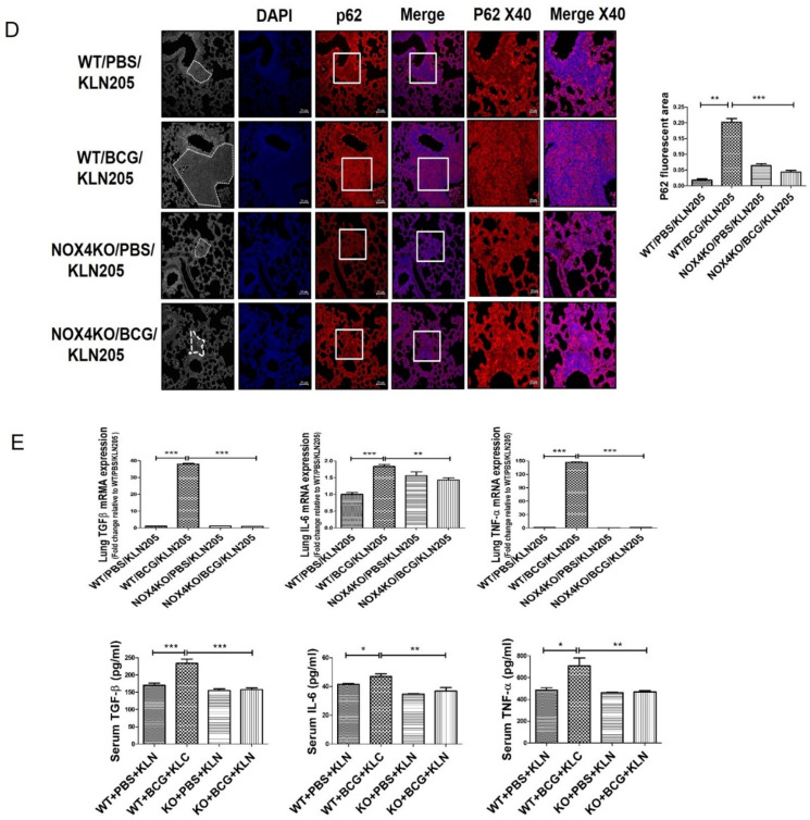
NOX4 mediates BCG pleurisy-promoted increased metastatic potential of lung cancer cells: (A) C57BL/6 NOX4 WT and KO mice were administered an intrapleural injection of BCG (1 × 10⁶ CFUs BCG Pasteur in 100 μL of saline). Each mouse also received an intravenous injection of KLN205 mouse lung cancer cells (2 × 10⁵) at day 29. At day 43, lung tissues were harvested. Representative micrographs of Bouin staining of metastatic nodules are presented. (B) Histological images of lungs. H&E staining (50×, 200×) and Ki67 immunohistochemistry (400×). Ki-67-positive cells show nuclear brown staining. (C) Lung cancer tissue lysates from each mouse were used for Western blotting of P62, ATG5-ATG12, LC3II/LC3I and Snail. (D) Activation of p62 deposition is regulated by NOX4 in a BCG pleurisy lung cancer model. p62 was subjected to immunofluorescence staining. Immunofluorescence staining of p62 (red); DAPI staining (blue); Purple represents colocalization of DAPI staining and immunofluorescence of p62. (E) RT-PCR analysis of TGF-β, IL-6, and TNF-α mRNA levels in mouse lung tissue lysates. ELISA analysis of TGF-β, IL-6, and TNF-α levels in mouse sera. Eight mice were used in each group (n= 8 per group). *** p < 0.001, ** p < 0.01, * p < 0.05.


