Figure 4.
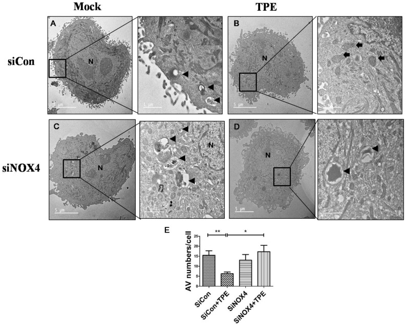
Loss of NOX4 increases autophagic vesicle abundance in A549 cells with TPE (tuberculous pleural effusion):A549 cells were transfected with siCon or siRNA targeting NOX4 (SiNOX4) for 1 day followed by treatment with or without TPE for 48 h. (A) siCon (negative control) group; (B) siCon+ TPE group; (C) siNOX4 group; (D) siNOX4+ TPE group. Abundant autophagic vesicles (AVs) were observed in A549 cells but few AVs were observed by transmission electron microscopy following treatment with TPE. Black arrowheads point to AVs including digested material. Black arrows indicate mitochondria. (E) The number of autophagic vesicles (AV) in each cell was counted with 5 cells in each sample, respectively. Data indicate the results of three experiments. ** p < 0.01, * p < 0.05.

