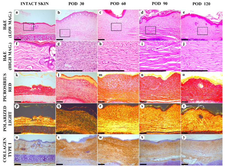
Figure 2.
Histological examination of the intact skin and experimental scars. (a–j) hematoxylin and eosin (H&E) staining; (k–t) Picrosirius red (PSR) staining; (u–y) immunohistochemistry (IHC) staining for collagen type I. Columns depict the studied groups (intact skin and experimental scars on PODs 30, 60. 90 and 120). Images were taken in bright-field (a–o,u–y) or polarized light (p–t) illumination. Scale bars are 200 µm.

