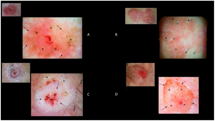
Figure 4.
(A): Clinical image of a 12 × 8 mm nonpigmented Bowen’s disease of the chest with dermoscopic features of dotted and glomerular vessels (↑) and scaly white-to-yellow surfaces (×). (B): Clinical image of a 30 × 12 mm pigmented Bowen’s disease of the leg with dermoscopic features of brown to gray globules/dots (↑) and structureless pigmentation (×) and dotted and glomerular vessels (>). (C): Clinical image of a 14 × 11 mm well differentiated cSCC of the scalp with dermoscopic features of keratin/scales (↑), blood spots (>), white structureless areas (×), and ulcerations (∞). (D) Clinical image of a 7 × 6 mm poorly differentiated cSCC of the ear with dermoscopic features of hairpin and linear-irregular vessels (↑) and ulcerations (×).

