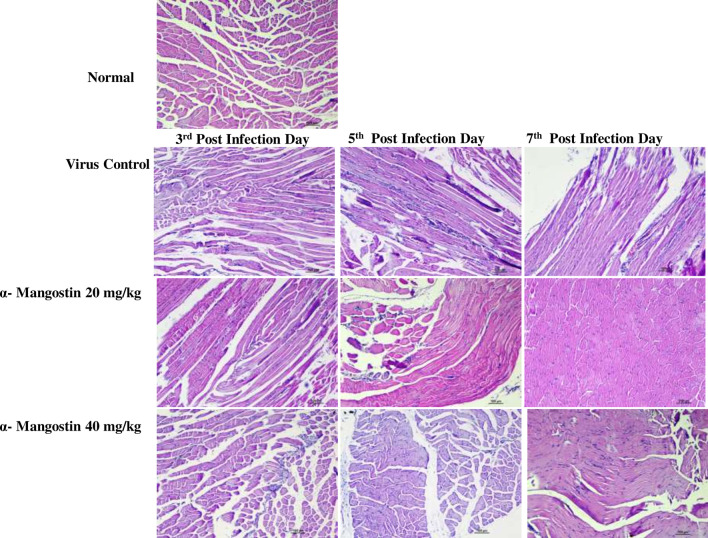
Fig. 4.
Histopathological changes in mouse muscle tissues after chikungunya infection and α-mangostin treatment. C57BL/6 mice were infected with CHIKV. Hematoxylin and eosin-stained tissue sections were screened to investigate the therapeutic effects of α-mangostin treatment in CHIKV infected mice. PBS injected mice showed normal cellular organization. CHIKV infected muscles showed marked muscle degeneration, atrophy, MNC infiltration and edema (at day 3, 5 and day 7). Treatment with low dose α-Mangostin showed improvement in inflammatory signs in muscle tissue at 7th dpi compared to the VC group. High dose α-Mangostin treated mice muscle tissues showed the regeneration after treatment

