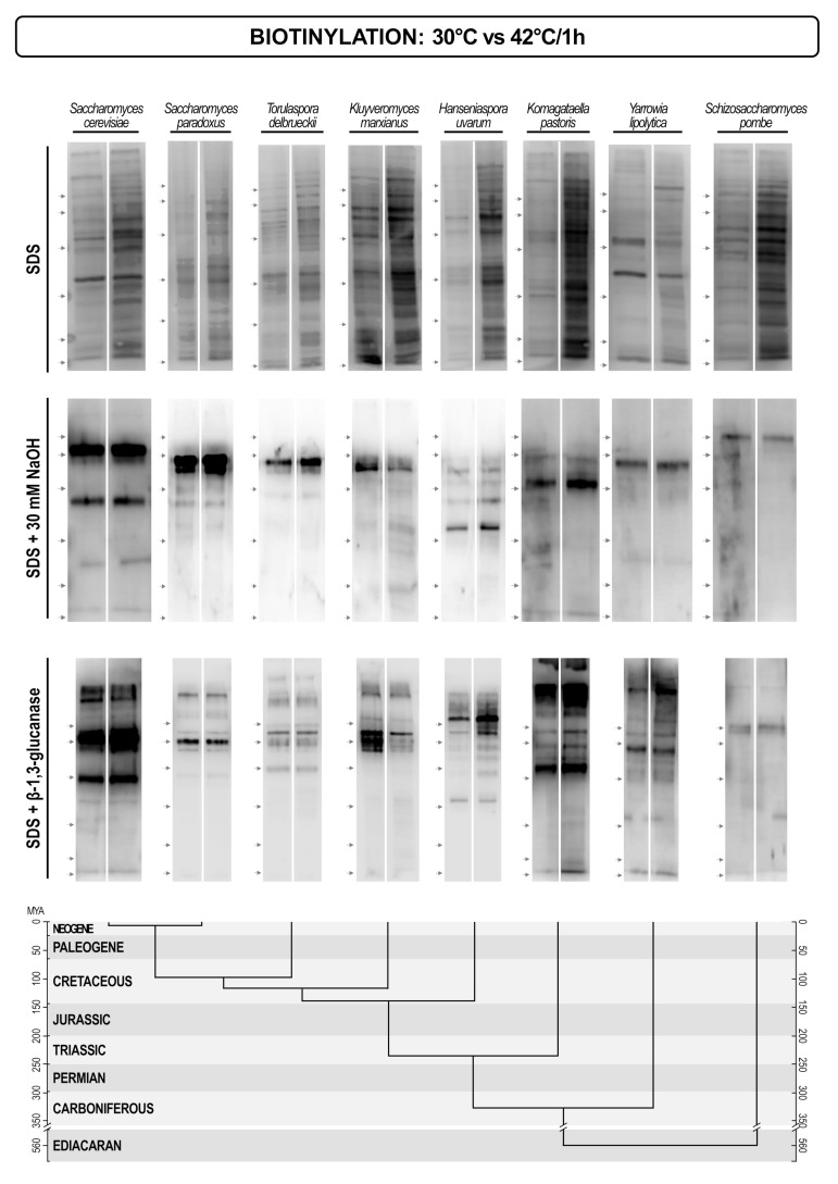
Figure 7.
Streptavidin/biotin blots of cell wall proteins of different yeasts extracted by hot SDS (non-covalently attached proteins), alkalis (Pir proteins + Scw4), and glucanases (GPI-anchored proteins). Proteins were extracted from cell walls of yeasts grown at 30 °C (left lane), and then shifted to 42 °C for one hour (right lane). The lower part of the figure presents the evolutionary chronogram of each species. Position of protein molecular mass markers is indicated on the left of each blot with protein sizes identical to those in Figure 6.

