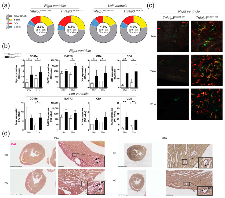
Figure 1.
Increased myocardial infiltration of dendritic cells (DCs) in the right ventricle (RV) of Tnfaip3DNGR1-KO mice. (a) Flow cytometry analysis for CD45+ cells (alive CD45+ cells), DCs (CD3-/CD19-, MHC-II+CD11c+), T cells (CD3+) and B cells (CD19+) in separately measured right and left ventricle cell suspensions from hearts of Tnfaip3DNGR1-WT/KO mice; (b) mRNA expression (relative to the glyceraldehyde-3-phosphate dehydrogenase (GAPDH) housekeeping gene) of DC markers CD11c and BATF3, and the T cell subset markers CD4 and CD8 in 24- and 31-week-old Tnfaip3DNGR1-KO and wild-type (WT) control mice; (c) whole mount analysis of right ventricle staining for YFP (green) and MHCII (red) in age 14-, 24- or 31-week-old Tnfaip3DNGR1-KO mice. Results are presented as mean values + standard deviations of 4–6 mice per group. * p < 0.05, ** p < 0.01; (d) Elastin van Gieson (EvG)-stained whole heart section histology of Tnfaip3DNGR1-WT and Tnfaip3DNGR1-KO mice for representative sections. Scale in left panels is 2.5 mm and in right panels 100 µm. Arrows indicate lymphocytes.

