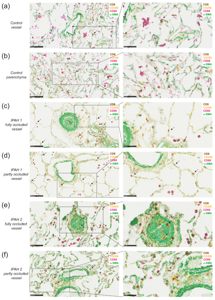
Figure 5.
DCs are in close proximity to CD8+ T cells around vessels and in parenchyma in lungs of idiopathic pulmonary arterial hypertension (IPAH) patients. (a,b) 4-plex chromogenic multiplex staining of DCs (CD206+, yellow), macrophages (CD68+, purple with or without CD206), T cells (CD8, DAB) and α-SMA (green) around a vessel and intraparenchymal (a) or in parenchyma (b) and the magnification of the indicated area (right) in the same healthy tissue from a smoking patient who was diagnosed with adenocarcinoma; (c,d) determination of DCs (CD206+CD68-), CD8+ T cells (CD8+), macrophages (CD68+) and vessels (α-SMA) around a fully occluded vessel (c) or partly occluded vessel (d) and the magnification of the indicated area (right) of the same IPAH patient with moderate remodeling; (e,f) determination of DCs, CD8+ T cells, macrophages and vessels around a fully occluded vessel (e) or partly occluded vessel (f) and the magnification of the indicated area (right) of the same a IPAH patient with extensive remodeling and immune cell infiltration. Data shown are representative for 6 IPAH patients. Sporadic CD206+CD68- cells with a morphology suggesting neutrophil identity were not regarded as DCs. Scale in left panels is 100 µm and of right panels 50 µm. Arrows indicate DC and CD8 co-localization

