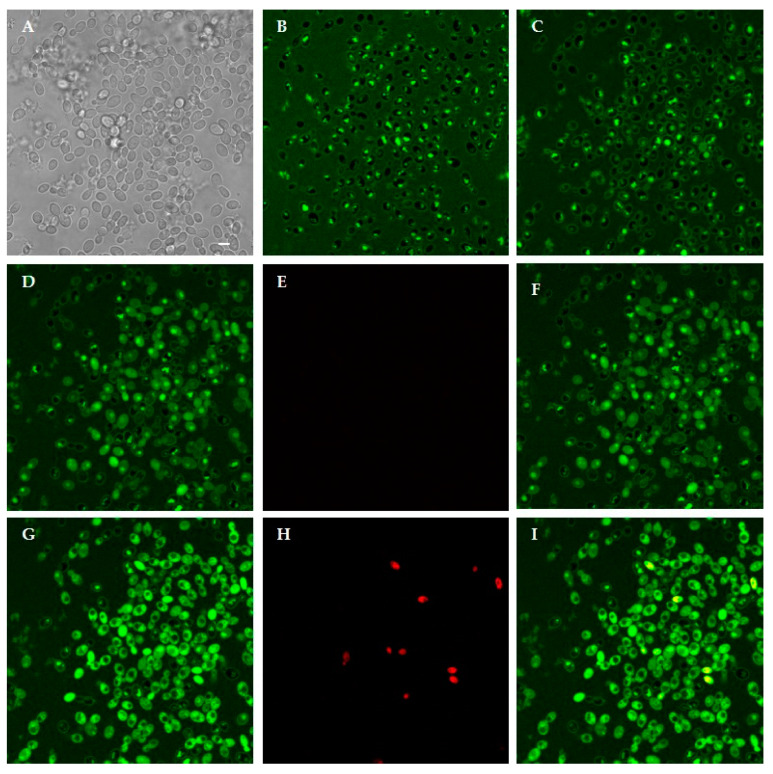
Figure 9.
Interaction between living Candida albicans cells and FITC-labeled H10S. Confocal microscopy images of yeast cells incubated for 10 min and 180 min with the labeled peptide are shown in (B,C), respectively. H10S was internalized in most yeast cells. In (A), the same field is shown in light transmission. Peptide internalization increased in viable cells after 240 min: (D) FITC; (E) PI; (F) merge of (D,E), eventually leading to cell death, as demonstrated by PI internalization at 360 min: (G) FITC; (H) PI; (I) merge of (G,H). Bar, 5 μm.

