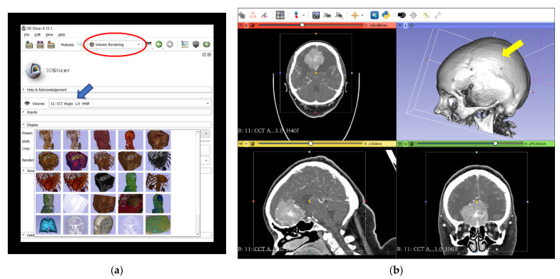
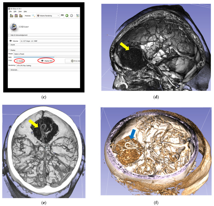
Figure 1.
Reconstruction process of 3D-VR images from 2D-CT modalities (including CTA) and completion of the final VR scene in the 3D Slicer. (a) Import of original CT and CTA data in an anonymized DICOM format into 3D Slicer software to create a patient-specific database and selection of “CCT Angio-Default” (blue arrow) in the volume rendering window (red circled). (b) Performance of 3D-VR reconstruction of a skull (yellow arrow) in the volume rendering window. (c) Activation of the ROI function (red circled), which enables visualization of the meningioma and relevant vascular anatomy from different perspectives by partial omission of skull bones. (d) Lateral aspect of the meningioma (yellow arrow) and relevant vascular anatomy, simplified by using the ROI function. (e) Superior aspect of the meningioma (yellow arrow). (f) Superior lateral aspect of the meningioma (blue arrow) with different color displays. 2D, two-dimensional; 3D, three-dimensional; CT, computed tomography; CCT, cranial computed tomography; CTA, computed tomography angiography; DICOM, digital imaging and communications in medicine; ROI, regions of interest; VR, virtual reality.


