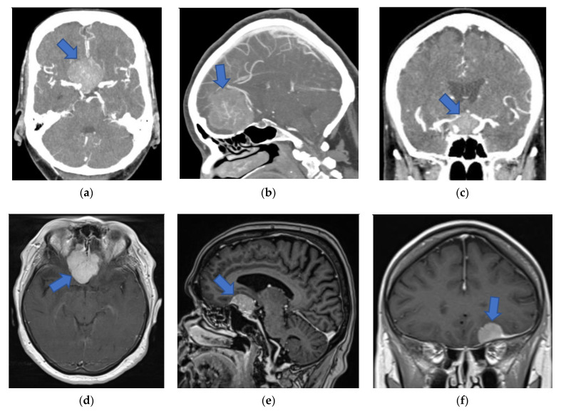
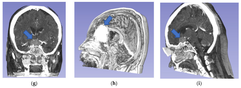
Figure 2.
Preoperative 2D and screen 3D images (from CT and MRI modalities) of patients with anterior skull base meningiomas (blue arrows). (a) Axial 2D-CTA image presenting planum sphenoidale meningioma. (b) Sagittal 2D-CTA image presenting olfactory groove meningioma. (c) Coronal 2D-CTA image presenting tuberculum sellae meningioma. (d) Axial 2D-MRI image presenting olfactory groove meningioma. (e) Sagittal 2D-MRI image presenting anterior clinoidal meningioma. (f) Coronal 2D-MRI image presenting frontobasal meningioma. (g) Coronal screen 3D-CT image presenting tuberculum sellae meningioma. (h) Sagittal screen 3D-MRI image presenting olfactory groove meningioma. (i) Sagittal screen 3D-CT image presenting anterior clinoidal meningioma. 2D, two-dimensional; 3D, three-dimensional; CT, computed tomography; CTA, computed tomography angiography; MRI, magnetic resonance imaging.


